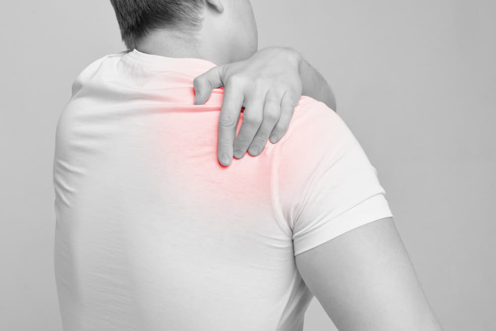
Shoulder instability and treatment

The glenohumeral joint (shoulder) joint is a ball and socket joint. The ‘ball’ (humeral head) sits inside the socket (glenoid). It facilitates a large range of upper limb motion that is essential for undertaking most activities of daily living. The downside of this large range of motion is the potential for instability. Ligaments, joint capsule, labrum and tendons help provide additional stability to the joint.
What is glenohumeral instability?
Glenohumeral instability is defined as an inability to maintain the humeral head centred
in the glenoid fossa. The ball usually slips off the front of the socket, and in doing so it damages the ligaments and joint capsule at the front of the shoulder. As the ball recoils back and hits the front of the socket, a dent can be left in the back of the ball.
What causes glenohumeral instability?
Glenohumeral instability can occur during a fall or in a sporting collision where the arm is twisted at an awkward angle. It can also occur after a direct blow to the shoulder. Patients report feeling pain, discomfort and/or a dead arm sensation.
What are the different types of glenohumeral instability?
Instability of the shoulder can be in one direction e.g., anterior instability (out the front), posterior instability (out the back) or in more than one direction (multidirectional instability).
Multidirectional instability usually occurs in patients with ligamentous laxity (very flexible, ‘double jointed’).
Anterior instability is the most common, followed by multidirectional, then posterior.
How is glenohumeral instability diagnosed?
The diagnosis is often clear from the history.
Your doctor will perform a range of physical examination manoeuvres.
Radiographs (x-rays) of the shoulder will be taken to ensure that there is no gross bone abnormality.
Magnetic resonance imaging (MRI) with injection of dye into shoulder (arthrogram) will be performed to have a better view of the labrum and ligaments around the shoulder
What treatment options are available?
In the acute setting, the dislocated shoulder needs to be put back in the correct position. This is usually done in a hospital emergency department.
Once the shoulder joint is reduced, treatment usually involves a gentle rehabilitation of the shoulder with a physiotherapist.
The risk of re-dislocation will influence whether surgery is recommended or not. Younger men tend to be at a higher risk of redislocation.
Arthroscopic (keyhole) surgery may be recommended to repair the torn labrum. It there is bone loss on the glenoid after the dislocations, a piece of adjacent bone may be transferred to the front of the socket to provide stability (Latarjet procedure).
After the surgery, a patient will need to undergo rehabilitation. This improves the strength and control of the small muscles (dynamic stabilisers) that help to keep the ball connected to the socket.
Shoulder surgery: LMcG Orthopaedics
Shoulder instability can cause pain and affect your way of life. At LMcG Orthopaedics, Doctor Lorcan McGonagle provides a range of orthopaedic surgeries and treatments. Dr McGonagle’s specialties include knees, shoulder, elbow, wrist and hand, hip, foot and ankle.
If you have a question for Dr McGonagle and his Geraldton team, please get in touch with the clinic today on 08 9921 4847. LMcG Orthopaedics is in Suite 6, St John of God Specialist Centre, 12 Hermitage Street, Geraldton. Click here to make an appointment.
